


Research, Learning and Innovation

Research allows our hospital to pioneer life-changing and life-saving care and treatments. To champion our Trust’s research ambitions, we dedicate £1m every year to support high-quality research

Hysteroscopic classification for endometrial polyps

Charitable funding has enabled research into the use of hysteroscopy, a convenient and accessible outpatient test, to detect the nature of endometrial polyps.  

Endometrial polyps are highly prevalent structural abnormalities found in the uterus. Removal of these polyps has traditionally been recommended to treat symptoms of abnormal uterine bleeding or infertility. 

Data is currently lacking to support the idea a polypectomy – a highly invasive and often painful treatment which regularly requires a general anaesthetic and long recovery periods – is effective in alleviating symptoms or offering a solution. 

However, the need to remove polyps for further examination to rule out potential malignant or premalignant tissue is still a necessity, which is why this research has been developed to find a less invasive, safer way of diagnosing the nature of endometrial polyps.  

Currently, to diagnose endometrial polyps, a hysteroscopy is conducted. While its use for diagnosis is foolproof, its efficiency for determining the nature of a polyp – whether it’s likely to be benign or not – has received little attention. This upcoming research will look at the accuracy and reproducibility of using a hysteroscopy to evaluate the nature of polyps which can then go on to inform clinical decisions

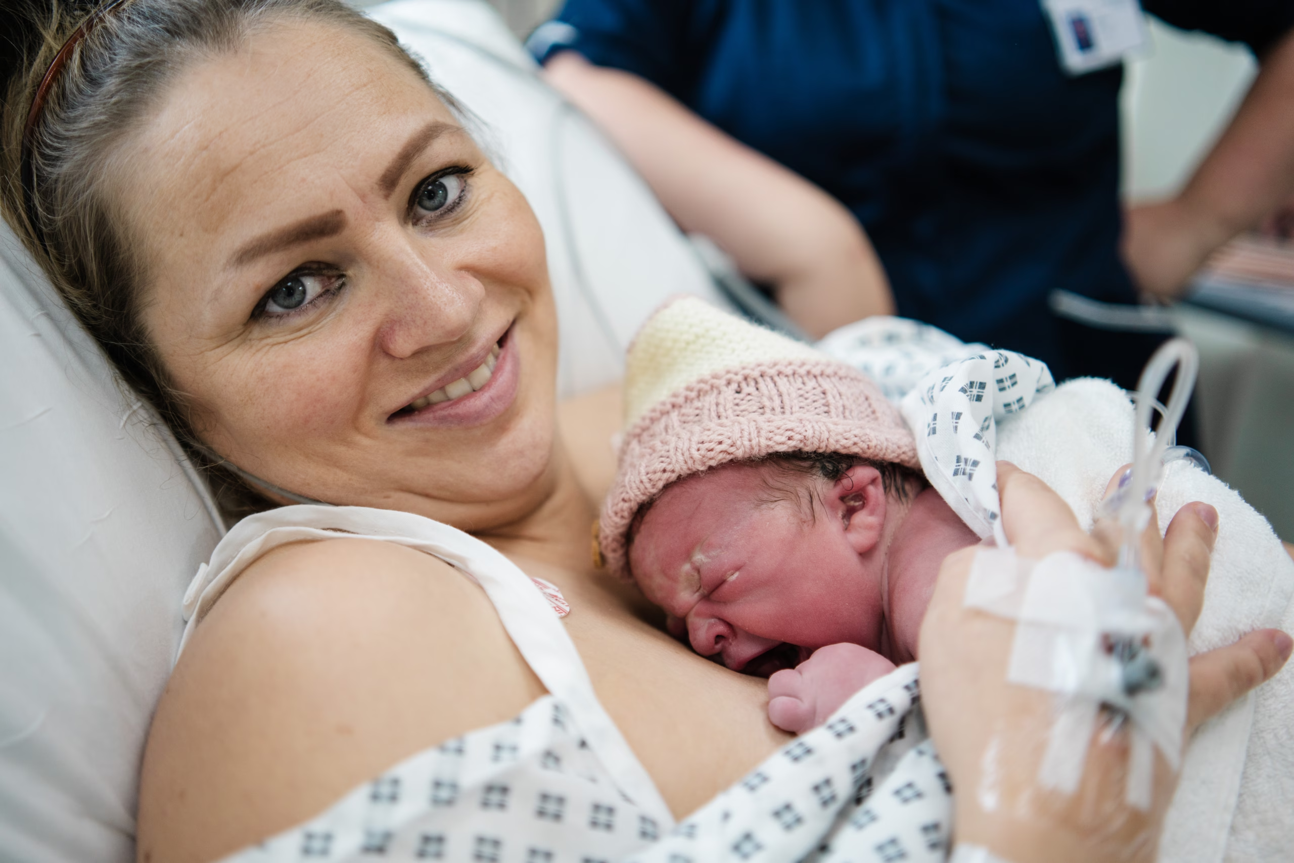
Mum Sarah Greenow holding baby, Monty

Placental characteristics within monochorionic twin pregnancies

Twin pregnancies which share a placenta, termed monochorionic, are at an increased risk of foetal complications, such as selective intrauterine growth restriction (where one twin grows more than the other) and twin-to-twin transfusion syndrome (where blood passes unequally between twins sharing a placenta).

The placenta is connected to the developing babies by tubelike structures called umbilical cords, which provide oxygen and nutrients. Each baby shares a portion of the placenta, allowing each one to grow. While the function of the placenta is intended to be the same for each pregnancy, each placenta develops slightly differently. 

This study, funded by a legacy left to our charity, looks to answer an important question – whether differences in the placental anatomy, such as the umbilical cord location, play a role in how twin pregnancies develop.  

Understanding if certain placental features cause some pregnancies to be uncomplicated and others to develop complications will help improve the way we care for women with twin pregnancies in the future. 

This could be by enhancing patient counselling for those who suffered complications during pregnancy, potentially providing clarity as to why certain outcomes occurred. Additionally, if study results demonstrate significant associations, then this could impact how we conduct future monochorionic antenatal surveillance, such as incorporating additional prenatal imaging.